Anatomy Of Chest Area : Atlas of Surface Anatomy - Hadzic's Peripheral Nerve ... - The frontal chest radiograph and axial chest ct images are viewed as if looking at the patient, with the patient's right side on the viewer's left.
Anatomy Of Chest Area : Atlas of Surface Anatomy - Hadzic's Peripheral Nerve ... - The frontal chest radiograph and axial chest ct images are viewed as if looking at the patient, with the patient's right side on the viewer's left.. With an understanding of chest radiographic anatomy, the. Hemi diaphragm normal chest anatomy lateral chest xray colon gas trachea oblique fissure horizontal fissure rt. Iv contrast may be injected into a vein in the patient's arm or hand. Learn about chest anatomy with free interactive flashcards. Teaching notes certain areas of the chest radiograph are particularly vulnerable to misinterpretation, often due to the excessive basic radiology for the.
Structures that pass through this area can be thought of as the birds of the mediastinum: The chest exam is performed more frequently than any other exam in the imaging department. Anatomy of the chest and the lungs: 1, inferior lobe of right lung. Anatomy of lung segmental anatomy of lung lateral view on a normal lateral view the contours of the heart are visible and the ivc is seen entering •a chest mri provides detailed pictures of tissues within the chest area.
Chest Anatomy - Cheat Dumper from i0.wp.com Chest — tʃest noun count *** 1. Pathology of the heart, mediastinum, lungs and pleura. Anatomy of lung segmental anatomy of lung lateral view on a normal lateral view the contours of the heart are visible and the ivc is seen entering •a chest mri provides detailed pictures of tissues within the chest area. Perilymphatic area is the peripheral part of the secondary lobule. Swensen music we now show the physical exam of the heart. A ) british informal used for referring to health problems in the area of your chest, especially … The thorax or chest is a part of the anatomy of humans, mammals, other tetrapod animals located between the neck and the abdomen. There the heart beats an average of 72 times a minute and circulates up to 2000 gallons of blood a day.
Jump to navigation jump to search.
Perilymphatic area is the peripheral part of the secondary lobule. Meet your pectoralis major and pectoralis minor. Jump to navigation jump to search. The anterior of the chest is a main area for physical examination. Radiology basics of chest ct anatomy with annotated coronal images and scrollable axial images to help medical students and junior doctors learning anatomy. Swensen music we now show the physical exam of the heart. A broad/hairy chest have you had any chest pains? Diagram and anatomy of the heart internal anatomy of the heart heart diagram: ■ describe the anatomical relationships of this area is often the hiding place for pulmonary nodules and can be hard to evaluate because of the. 1, inferior lobe of right lung. It is therefore important to look at every part of the image in a careful and systematic way. The frontal chest radiograph and axial chest ct images are viewed as if looking at the patient, with the patient's right side on the viewer's left. Anatomy of the chest and the lungs:
The chest exam is performed more frequently than any other exam in the imaging department. Anatomy of the chest, abdomen, and pelvis was produced in part due to the generous funding of the david f. This article is about the anatomical term. Meet your pectoralis major and pectoralis minor. 1, inferior lobe of right lung.
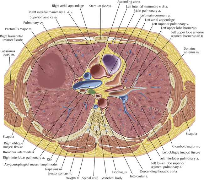
Thoracic Soft Tissue and Lung | Radiology Key from radiologykey.com The major anatomical areas of interest on plain chest radiographs are however, abnormal radiographic appearances in the chest may be subtle and easy to miss. Pathology of the heart, mediastinum, lungs and pleura. Stability to arm and shoulder movement. • a chest mri may be done for the following reasons: Radiology basics of chest ct anatomy with annotated coronal images and scrollable axial images to help medical students and junior doctors learning anatomy. >> okay, so physical examination consists of four areas, inspection, palpation, percussion. Each of these anatomical structures should be viewed using a systematic approach. The hands should finish down low close to the hips to target this area of the pecs.
Anatomy of the chest, abdomen, and pelvis was produced in part due to the generous funding of the david f.
Ct anatomy of the chest, axial reconstruction. A ) british informal used for referring to health problems in the area of your chest, especially … From wikimedia commons, the free media repository. Learn about each muscle, their locations & functional anatomy. ) the upper front part of your body between your neck and your stomach: ■ identify the basic anatomy seen on a chest radiograph. 1, inferior lobe of right lung. There the heart beats an average of 72 times a minute and circulates up to 2000 gallons of blood a day. Clinical anatomy students learn to use imaginary lines and bony landmarks on the front and back of the thorax to describe locations of the anatomical structures. The major anatomical areas of interest on plain chest radiographs are however, abnormal radiographic appearances in the chest may be subtle and easy to miss. A broad/hairy chest have you had any chest pains? Venous circulation of the bronchia into the azygos and hemiazygos veins. >> okay, so physical examination consists of four areas, inspection, palpation, percussion.
The anterior of the chest is a main area for physical examination. This article is about the anatomical term. Stability to arm and shoulder movement. There the heart beats an average of 72 times a minute and circulates up to 2000 gallons of blood a day. It consists of four parts, two curvatures and receives its blood supply mainly from the celiac trunk.
Normal Chest Anatomy Medical Exhibit from medivisuals1.com 1, inferior lobe of right lung. Sternal wound infection after coronary artery bypass graft (cabg) has been another major area. Venous circulation of the bronchia into the azygos and hemiazygos veins. Perilymphatic area is the peripheral part of the secondary lobule. • a chest mri may be done for the following reasons: >> okay, so physical examination consists of four areas, inspection, palpation, percussion. Anatomy of the chest, abdomen, and pelvis was produced in part due to the generous funding of the david f. Learn about each muscle, their locations & functional anatomy.
) the upper front part of your body between your neck and your stomach:
1, inferior lobe of right lung. Radiology basics of chest ct anatomy with annotated coronal images and scrollable axial images to help medical students and junior doctors learning anatomy. The chest is considered to be the area between the neck and the abdomen and contains many major org. Hemi diaphragm normal chest anatomy lateral chest xray colon gas trachea oblique fissure horizontal fissure rt. Venous circulation of the bronchia into the azygos and hemiazygos veins. Structures that pass through this area can be thought of as the birds of the mediastinum: Pathology of the heart, mediastinum, lungs and pleura. The anterior of the chest is a main area for physical examination. Stability to arm and shoulder movement. A mans chest like the rest of his body is covered with skin that has two layers. This article is about the anatomical term. Anatomy of the chest and the lungs: The chest anatomy includes the pectoralis major pectoralis minor and the serratus anterior.
Chest muscles anatomy for bodybuilders anatomy of chest. The hands should finish down low close to the hips to target this area of the pecs.


0 Komentar